
By CareMotion · Modalities & Technology
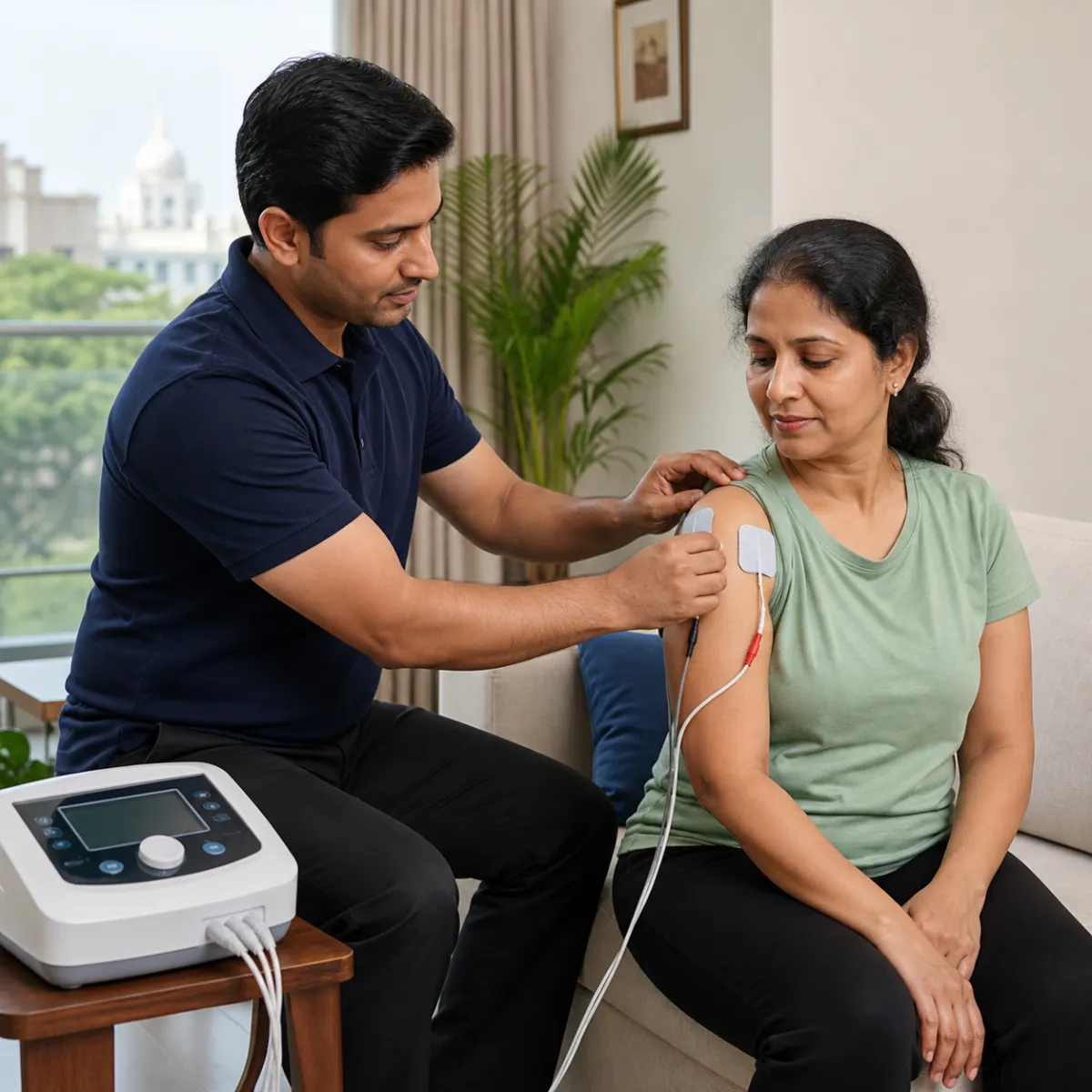
What is Therapeutic Ultrasound (UST) in Physical Therapy?
Ultrasound Therapy (UST) uses high-frequency mechanical sound waves (typically 1 MHz or 3 MHz) that lie far beyond human audible perception. Delivered through a piezoelectric crystal within a specialized metal applicator, sound waves transmit mechanical energy into biological tissues.
UST is completely different from diagnostic medical ultrasound used for imaging. In physiotherapy, ultrasound is applied therapeutically to generate micro-acoustic streaming, cellular permeability enhancement, and thermal deep tissue heating.
- 1 MHz frequency penetrates deeply into tissues 3-5 cm beneath the skin (ideal for hips, quads, and lumbar discs).
- 3 MHz frequency targets superficial tissues 1-2 cm deep (ideal for tennis elbow, Achilles tendon, and wrist ligaments).
- Acoustic coupling gel is essential to eliminate air gaps between the applicator head and skin.
Thermal vs. Non-Thermal Effects on Tissue Repair
Therapeutic ultrasound produces both thermal and non-thermal cellular effects depending on whether it is delivered in continuous or pulsed duty cycles. Continuous ultrasound increases local tissue temperature, relaxing contracted collagen fibers in chronic scars and stiff joints.
Pulsed ultrasound (such as 20% duty cycle) delivers mechanical acoustic streaming and stable cavitation without heating, making it suitable for acute tendon injuries, ligament sprains, and early soft tissue trauma.
- Increases local blood flow and lymphatic drainage in chronically stiff tendons.
- Enhances cell membrane permeability, stimulating fibroblast collagen synthesis.
- Softens dense post-surgical scar adhesions and fibrous joint capsular tightness.
Clinical Safety and Best Practices at Home
The applicator head must be kept continuously moving in circular or stroking patterns to prevent acoustic standing waves and localized periosteal bone ache. A licensed clinician monitors power intensity (measured in Watts per square centimeter, W/cm²).
Ultrasound therapy is never applied over malignant tumors, developing epiphyseal growth plates in children, acute infection sites, or directly over the spinal cord after a laminectomy.
- Treatment duration is calibrated based on the effective radiating area (usually 5-8 minutes per zone).
- Best combined with immediate passive stretching while tissues remain warmed and pliable.
- Always verified by a qualified physiotherapist with appropriate equipment calibration.
General information to help you plan a visit. Individual assessment and your treating team's advice guide rehabilitation decisions.